Enhance your clinical skills through palpation, inspection and movement
With Instructor Jamie Bender L.Ac., DAOM
Friday, January 9, 2026, 9:00-6:00, at ACCHS, 1600 Broadway, Oakland CA
Precise knowledge of clinical anatomy and kinesiology, and orthopedic/myofascial palpation and inspection, and movement analysis skills, are all essential foundations for diagnosis, and for determining where--and where not--to needle.
This unique class prepares students to get the most from the Low Back, Core & Pelvic Girdle module & Review/Practicum Lab.
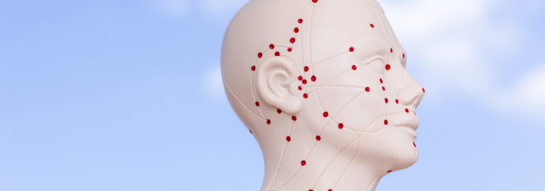
Clinical anatomy and the jing-jin ("sinew meridians" or myofascial tracts)
- We will improve our abilities to accurately locate key bony landmarks, muscles, tendons, joints, neural and vascular tissues, through palpation on ourselves and each other, and through review of clinical anatomy.
- Through palpation, observation and movement exercises, we will explore functions of key muscles and their jing-jin associations, as well as functional vs. dysfunctional movement patterns.
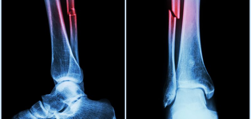
- We will review safety considerations, including needling angle and depth, to avoid injuring the many critical structures in this body region.
Enhanced orthopedic palpation and inspection skills
- We will enhance our abilities to feel different tissue types and layers: skin, fascia, muscle, nerve, blood vessel, and bone, with both our hands and needle-tip sensation.
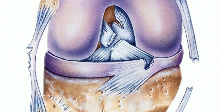
- We will practice inspection and palpation for tissue abnormalities including myofascial trigger points, tendinopathies and joint disorders.
Review of anatomical structure and kinesiologic function
- Bony landmarks. Also important to know which muscles attach to them, if applicable.
- 11th and 12th ribs
- Spinous processes of T 1 to L 5, lumbosacral junction
- Sacro-iliac joint line, sacral foramina
- Ilium, including crest, PSIS and ASIS
- Greater trochanter
- Myofascial structures that move and stabilize the lumbosacral spine and pelvis. Also know attachments.
-
- Erector spinae group
- Quadratus lumborum
- Iliacus and psoas
- External obliques
- Rectus abdominus
- Gluteus maximus, medius, and minimus
- Piriformis
Deep anatomy: know locations relative to surface anatomy and other deep structures
- Lumbosacral Spine
-
-
- Intervertebral discs: nucleus pulposus, annulus fibrosis
- Posterior longitudinal ligament
- Neuroforamen
- Central canal
- Neural arch: laminae, pedicles
-
- Lumbosacral neurology
-
- Spinal cord
- Nerve roots L 1 through S 4
- Sciatic nerve
Functional anatomy and anatomical kinesiology: know attachments and/or functions of the following
- Lumbosacral neurology
-
-
- Dermatomes
- Myotomes
-
- Lumbosacral, gluteal and abdominal anatomical kinesiology
-
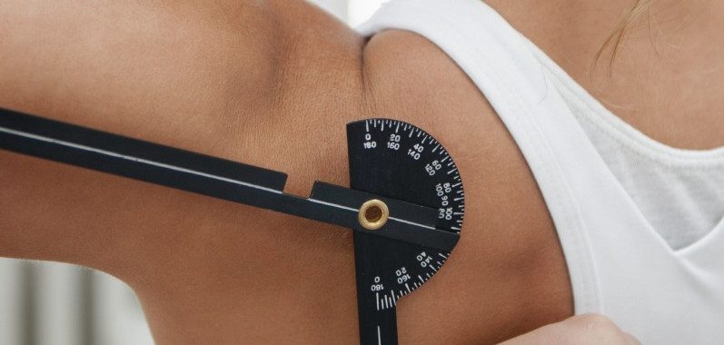
- Planes of lumbar motion
- Primary actions of the following muscles, when contracting unilaterally vs. bilaterally
- Erector spinae group
- Multifidi
- Quadratus lumborum
- Iliopsoas
- Abdominals: rectus, obliques, transversarius
- Gluteals: maximus, medius, minimus
- Piriformis